PET / CT Imaging
What is PET Imaging?
Positron Emission Tomography (PET) is a Nuclear Medicine
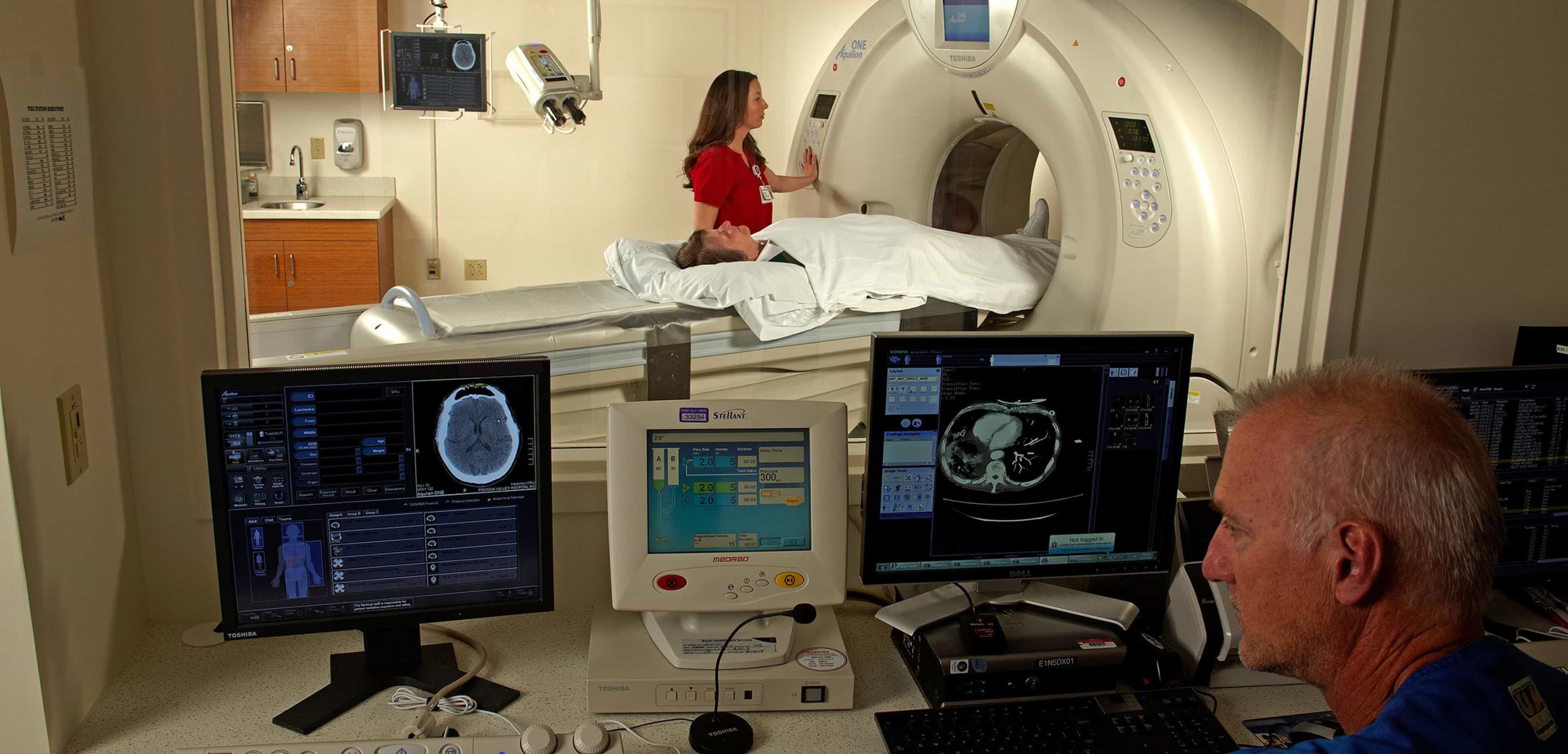
Imaging test currently provided by the PVHMC Radiology Department. PET scanners have been around in academic centers for decades, and due to improvements in the imaging technology and lower cost of the radio-pharmaceuticals they have now made their way into mainstream medical usage. PET scanners are being employed most frequently today in the diagnosis and staging of cancer, and are recognized by Medicare for reimbursement in selected cancers. From the outside, a PET scanner looks just like a CT scanner (see photo).
The radio-pharmaceutical used most often today in PET imaging looks at the metabolism of glucose in the body. A radioactive form of glucose is injected into the patient and is absorbed in the areas of the body actively metabolizing glucose. The radioactive emissions from the tissue metabolizing glucose exit the body and are picked-up electronically by the detector ring (the machine) around the patient, then sent to the computer, determining the site of origin. The computer then generates the images interpreted by the Physicians. PET imaging with radioactive glucose is of particular value in cancer patients because malignant cells often show higher rates of metabolism of sugar compared to the surrounding normal tissue, and therefore show up as areas of more intense glucose localization (hot spots).
